LASIK Surgery Frequently Asked Questions
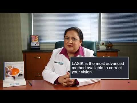
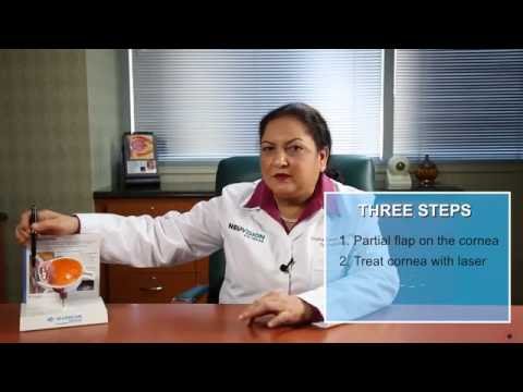
How does LASIK work?
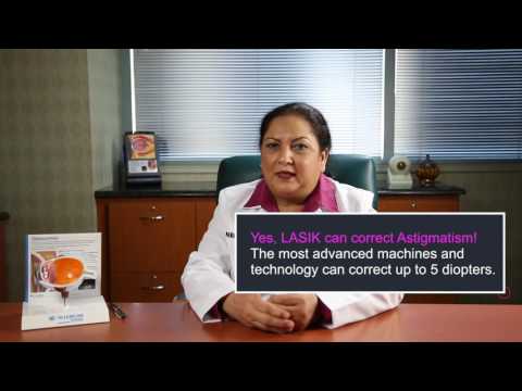
LASIK is a laser vision correction technique that reshapes the cornea to correct a wide range of vision disorders including nearsightedness, farsightedness and stigmatism.
Is LASIK safe?
Absolutely! The FDA approved LASIK for vision correction back in 1999. Today, it is one of the safest and most commonly performed procedures available.
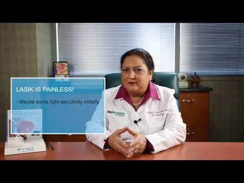
Does LASIK hurt?
No. The actual procedure is painless. However, you may feel some itchiness, dryness or pressure following your procedure. Our surgical team will give you eye drops and a prescription pain reliever for any potential discomfort.
Am I a candidate for LASIK surgery?
LASIK surgery isn’t right for everyone, and there are certain criteria a patient must meet to be considered a candidate including:
- A stable eyeglass or contact prescription
- Best corrected vision of at least 20/40
- Healthy cornea
- No active eye disease
- Over the age of 18 years old
Even if you aren’t a candidate for LASIK, there may be other vision correction options available to you. Only your eye doctor will know for certain.
How long is LASIK surgery?
The entire LASIK procedure generally takes less than 15 minutes per eye to perform. The laser portion of the vision correction surgery only takes a few seconds to complete. However, you should plan to be in the office on the day of your surgery for up to 2 hours.
How soon will I be able to see after LASIK surgery?
Recovery after LASIK surgery varies from patient to patient. Most patients return to normal activities one to two days after their procedure, but it may take up to 2 months for your vision to fully stabilize.
When will I be able to drive?
The overwhelming majority of LASIK patients regain legal driving vision or better within a day of their procedure, however that does not mean you are cleared to drive yourself home from your surgery. Plan to have a driver take you home and take you to your post-operative appointments. In most cases, patients are able to drive by the end of that day.
How long will my results last?
In most instances, laser vision correction is permanent, especially if your prescription was stable before surgery. During your consultation, your LASIK surgeon will discuss if there are any pre-existing conditions that will affect your results including genetics.
Will I need to wear glasses or contacts after LASIK surgery?
LASIK surgery permanently corrects vision problems, usually eliminating the need for glasses or contacts. However, LASIK doesn’t protect against age-related eye conditions or address certain refractive errors caused by cornea thickness. For this reason, some individuals who have had successful LASIK surgery may need glasses once again later in life. During your consultation, your LASIK surgeon will be able to address your concerns regarding glasses and contacts after LASIK.
Can I have both LASIK and cataract surgery?
Yes, you can have both LASIK and cataract surgery, but only in that specific order and most definitely not at the same time. If you’ve already had LASIK surgery, it’s possible you will develop cataract later in life. However, if you’ve already had cataract surgery, in most cases you are no longer a candidate for LASIK or other refractive surgeries. Thankfully, the insertion of an intraocular lens (IOL) after cataract surgery can accomplish similar vision correction results that you would obtain with LASIK surgery.
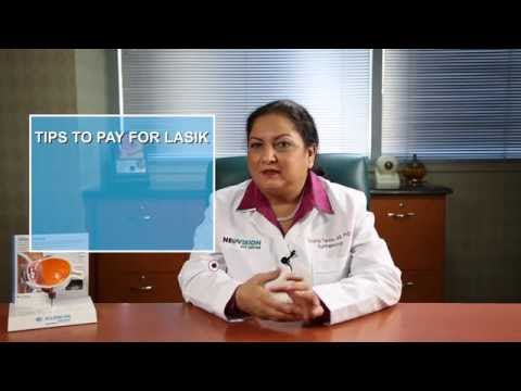
Do you offer financing?
At NeoVision Eye Center we understand that laser vision correction is an investment. We also believe that everyone has the right to better vision. For that reason, we offer many financing options ranging from short-term to long-term payment plans. Feel free to contact our staff to discuss which payment options best suit your budget and situation.